Facilitate the transition of projects from the laboratory to the clinic. Translational researchers seek to apply basic knowledge of cancer and bring the benefits of the new basic-level understandings to patients more quickly and efficiently. These grants are $600,000, three-year commitments
Co-funded by the Dick Vitale Gala, and WWE in honor of Connor’s Cure
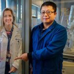
Dr. Jun Qi is a synthetic organic chemist and chemical biologist who has developed small molecules and pioneered anovel chemical strategy in which small molecule therapeutics can be designed to destroy specific proteins within a cell, as opposed to suppressing enzymatic function.Dr. Mariella Filbin is a physician scientist specializing in pediatric neuro-oncology with clinical and scientific interests converging upon pediatric brain cancers, in particular, diffuse intrinsic protein glioma (DIPG) which is universally fatal. Dr. Filbin has used patient-derived modelsto identify a potential DIPG-specific target for Dr. Qi’s protein degrader technology. They will work together to overcome challenges in childhood brain cancer treatment, such as toxicity and blood-brain-barrier (BBB) penetration.This exciting study has two broad objectives:
To define the mechanism by whichthe cancer dependent proteinis driving DIPG formation and growth;
To yield optimized drug compounds suitable for preclinical study and translation to clinical trials in DIPG.
By working together as team, Drs. Qi and Filbin will cultivate a symmetrical relationship in whichchemistry will be used to clarify the biology; and biology will be used to guide the small molecule design and development. By combining their complementary skill sets in chemistry, chemical biology and cancer biology, their joint efforts will result in the preclinical validation of eliminating the target genes and ideally the development of a clinical trial using this novel strategy for DIPG to achieve the bench-to-bedside translation of their research.
RAS is a gene that plays a major role in cancer. The three members of the RAS family are HRAS, NRAS, and KRAS. One of these genes is mutated in about 15% of cancers. The mutant form is hyperactive.
In pediatric solid tumors, RAS is mutated in about 1-3% of cancers and more often in rhabdomyosarcoma.
Inhibiting RAS activity has been a difficult task in cancer drug development. One type of drug, the farnesyl transferase inhibitors (FTI), were developed twenty years ago. Clinical trials using these drugs were disappointing. We now have a better understanding of how to select patients that will best respond to FTI.
Only mutant HRAS is dependent on the farnesyl transferase enzyme. So, FTI should work best in patients with HRAS mutant cancers.
In a clinical trial of patients with HRAS mutant head and neck cancer, patients were treated with tipifarnib, an FTI. Trial outcomes showed that patients’ tumors got smaller (responded).
We are now studying FTI in pediatric solid tumors. We want to know what adaptive events occur in the cell and whether these changes only occur in mutant HRAS tumors. We also want to learn how tumors may escape the anti-cancer effects of FTI.
Studying these changes and paths of resistance can help us develop more complete and lasting responses. Our study aims to address these issues to find effective treatments for patients with HRAS mutant cancer.
Co-funded by the Dick Vitale Gala, and WWE in honor of Connor’s Cure
DIPG is a universally fatal brain tumor that occurs in children. Thanks to extensive research, we now understand the biologic causes of DIPG, but no one has found an effective way to treat the disease. Patients receive radiation to slow the disease and relieve symptoms, but they almost all die within two years of diagnosis. We have found that a target known as GD2 is highly expressed on DIPG. GD2 can be targeted with an antibody that is FDA approved to treat another type of cancer. When the antibody finds its target, it recruits immune cells to “eat” the cancer cells. Here, we propose combining anti-GD2 with another antibody that stimulates the immune system to “eat” cancer cells (anti-CD47). Because antibodies cannot reach the brain when given in the blood, we will deliver these two antibodies by direct injection into the tumor. Our main goal is to test this approach in mouse models of DIPG to see if it is safe and effective. This will hopefully serve as the basis for a clinical trial for children with DIPG. We will also explore alternative and complementary ways to attack the tumors.
Co-funded by the Dick Vitale Gala, and WWE in honor of Connor’s Cure
Years of cancer research have shown that combining therapies virtually always works better than when therapies are used alone. Recently, medications have been discovered that change the way genes are turned on and off. At the same time, treatments have been developed that use the body’s own cells to find and attack cancer cells. Each of these treatments have been shown to work alone on specific cancers. Each has known limits. However, the combination has not been studied. Our project explores whether combining these treatments will improveour treatments for childhood cancer. We are especially interested in if combining these therapies will increase the success of cellular therapies. Our proposal initially studies one specific medication that is already approved for use in children. We will also evaluate a large group of possible medications. We expect that our results will quickly result in a clinical trial for children. In addition, it may lead to a new treatment approaches for many cancers.
The most difficult challenge in treatment management of brain tumor patients is the need to accurately identify if a suspicious lesion on a post-treatment MRI scan is a benign treatment-effect or a “true” cancer recurrence. Both radiation effects and tumor recurrence have similar clinical symptoms and appearances on routine MRI scans. Currently, a highly invasive brain biopsy is the only option for confirmation of disease presence. Each biopsy procedure costs $20,000-$50,000/patient. Further, over 15% of patients who undergo biopsy will get an incorrect diagnosis due to difficulty in sampling of reliable locations of the tumor. There is hence a need for non-invasive image techniques to reliably differentiate benign treatment effects from tumor in brain tumor patients. Our team has developed new image-based biomarkers that use routine MRI scans to differentiate between these two conditions with an accuracy of 92% on n>200 studies. We propose to validate our image-based biomarkers in a limited clinical trial to reliably sample locations of tumor recurrence from benign radiation effects. The clinical trial will be based on creation of a “GPS” map of the locations of tumor and benign radiation necrosis in the tumor using MRI scans. This GPS map will assist neurosurgeons in reliably identifying locations to biopsy from during surgery. The proposed project, when successful, will thus have significant implications in personalizing treatment decisions in brain tumors.
Funded by the Dick Vitale Pediatric Cancer Research Fund
There is a unique group of cancers that progress quickly during childhood due to faults in the mechanisms which repair damaged DNA. As a result, these childhood cancers have the highest number of DNA mutations (hypermutant) of all human cancers. Immunotherapy has demonstrated hopeful results in these patients. Yet, 50% of these cancers will progress after initial response to immunotherapy. This poses a significant problem. Adoptive cell therapy takes advantage of using immune cells to kill cancer cells. Cell therapy has shown promising responses in many adult cancers. This effect is greater when cell therapy is used in combination with prior immunotherapy treatment. Our research team has developed new mouse models that successfully mimic these childhood brain cancers. One of the aims of our research project is to use these mouse models to study the role of cell therapy. We will determine overall survival and response to therapy. We aim to prove the feasibility of expanding childhood immune cells as a proof of concept through the use of our International Consortium. We will use complex computer software and genomic tools. These methods will provide a thorough review of immune cells. We will be able to predict which patients would benefit from cell therapy. This project will increase knowledge in this research area. In addition, it will answer important questions which will lead to improved patient outcomes and treatment options. Most importantly, this project will lead to the first-ever childhood cell therapy clinical trial.
Brain tumors cause the most cancer deaths in children. A tumor known as medulloblastoma (MB) is the most common type of childhood brain cancer. Children die of MB because the cancer spreads through the brain. New information indicates that some MB cells may first go into the bloodstream before spreading to the brain and forming new tumors. Cancer cells in the bloodstream are called “circulating tumor cells” (CTCs). We recently developed a tool called Cluster-Chip that can detect CTCs in the blood and remove them so that they can be studied. Using our Cluster-Chip tool, we want to see how often CTCs are found in the blood, and in which MB patients we find them in. Next, we want to see exactly what CTCs look like, what they are made of, and if they are different from the rest of the brain tumor. Finally, we want to see whether the number, the appearance or the make-up of CTCs in the blood can tell us if the tumor will go on to spread to the brain and if the patient will die of their disease. We will study 25 patients with MB and collect their blood at different times throughout their treatment. This information will help us to understand how MB cancer spreads and how to better treat MB tumor spread.
Funded by the Constellation Gold Network Distributors in honor of the Dick Vitale Pediatric Cancer Research Fund
Philadelphia chromosome-like acute lymphoblastic leukemia (Ph-like ALL) is a common cancer in children and adults that does not respond well to regular chemotherapy medicines and often comes back. We found in earlier studies that Ph-like ALL has ‘miswired’ signaling networks inside its cells. These networks seem to be very sensitive to targeted medicines called kinase inhibitors. We are now testing one of these inhibitor medicines with chemotherapy in children with Ph-like ALL in a clinical trial, but we do not yet know if adding this new medication will be better than regular chemotherapy by itself. We will study leukemia cells from patients treated on this clinical trial to try to answer this question. We will also use specialized mouse models made from the children’s leukemia cells to understand what other miswired networks happen in Ph-like ALL and could be attacked by new medicines. These laboratory studies will help us to learn if using several inhibitor medicines together could be even better than current chemotherapy. If this is the case, then we will then hope to test this new treatment idea in children with Ph-like ALL in future clinical trials.
Vintner Grant funded by the V Foundation Wine Celebration in honor of Joe and Pat Harbison
DNA, which stores all of our genetic information, is constantly being damaged by environmental sources such as sunlight or from products of normal processes within each cell. If unrepaired, DNA damage may result in mistakes, which can lead to cancer. We study human cells from patients who do not have the full capacity to repair the DNA due to a genetic disease called Fanconi anemia. They are predisposed to the development of cancers including those of head and neck. We propose to determine how cancers develop in this group of patients by identifying all the permanent changes that occur in Fanconi anemia tumors and to study how these changes lead to cancer development. We also want to take advantage of these changes to find better treatments for head and neck cancers. For our work, we use patient tumor samples and mouse models of cancer. In addition to all of the tools we currently have at our disposal, we aim to develop new ones including patient tumor samples that can be grown in the mouse and can be shared across laboratories. Our studies have the potential to help with prevention, early detection, and treatment of head and neck cancers.
A standard treatment for bladder cancer that has invaded into the muscle layer of the bladder is to first give chemotherapy medication for several months and then surgically remove the bladder. Surgical removal of the bladder is a major operation and is associated with a potential risks. Also, because the bladder is where urine is stored in the body, when the bladder is surgically removed, the urine has to exit the body differently. For many patients, this means that the urine will be drained into a bag outside of the body called a urostomy. When chemotherapy medication is given through a vein for several months prior to surgery to remove the bladder, sometimes there is no more cancer in the bladder specimen when it is taken out of the body and inspected in the laboratory. If we could identify which patients might have their bladder cancer eliminated with chemotherapy medication alone, this could mean that some patients may be cured without having their bladder removed. We are testing whether given chemotherapy together with immunotherapy, medication to enhance the body’s immune system to fight cancer, is better at completely eliminating cancer in the bladder and also testing whether we can identify patients that are the best candidates for this approach by studying several features of an individual patient’s cancer before and after treatment. If our work is successful, we hope to be able to select patients who can have their bladder cancer cured with the combination of chemotherapy and immunotherapy without requiring surgical removal of their bladder.